Monday, March 3

CELLS - CHLOROPLASTS #12

Chloroplasts - Show me the Green


Structure of a chloroplastChloroplasts are the food producers of the cell. They are only found in plant cells and some protists. Animal cells do not have chloroplasts. Every greenplant you see is working to convert the energy of the sun into sugars. Plants are the basis of all life on Earth. They create sugars, and the byproduct of that process is the oxygen that we breathe. That process happens in the chloroplast. Mitochondria work in the opposite direction and break down the sugars and nutrients that the cell receives. 

Special Structures

Structure of thylakoid stackWe'll hit the high points for the structure of a chloroplast. Two membranes contain and protect the inner parts of the chloroplast. The stroma is an area inside of the chloroplast where reactions occur and starches (sugars) are created. Onethylakoid stack is called a granum. The thylakoids have chlorophyll molecules on their surface. That chlorophyll uses sunlight to create sugars. The stacks of sacs are connected by stromal lamellae. The lamellae act like the skeleton of the chloroplast, keeping all of the sacs a safe distance from each other and maximizing the efficiency of the organelle. 

Making Food
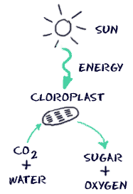
Super basic process of photosynthesisThe purpose of the chloroplast is to make sugars and starches. They use a process called photosynthesis to get the job done. Photosynthesis is the process of a plant taking energy from the Sun and creating sugars. When the energy from the Sun hits a chloroplast, chlorophyll uses that energy to combine carbon dioxide (CO2) and water (H2O). The molecular reactions create sugar and oxygen (O2). Plants and animals then use the sugars (glucose) for food and energy. Animals also use the oxygen to breathe. 

Different Chlorophyll Molecules

We said that chlorophyll molecules sit on the outside of the thylakoid sacs. Not all chlorophyll is the same. Three types of chlorophyll can complete photosynthesis. There are even molecules other than chlorophyll that are photosynthetic. One day you might hear about carotenoidsphycocyanin (bacteria), phycoerythrin (algae), andfucoxanthin (brown algae). While those compounds might complete photosynthesis, they are not all green or the same structure as chlorophyll. 

CHLOROPLAST - QUIZ

CELLS - MITOCHONDRIA #11


Mitochondria - Turning on the Powerhouse


Mitochondria are known as the powerhouses of the cell. They are organelles that act like a digestive system that takes in nutrients, breaks them down, and creates energy for the cell. The process of creating cell energy is known as cellular respiration. Most of the chemical reactions involved in cellular respiration happen in the mitochondria. A mitochondrion is shaped perfectly to maximize its efforts.

Mitochondria are very small organelles. You might find cells with several thousand mitochondria. The number depends on what the cell needs to do. If the purpose of the cell is to transmit nerve impulses, there will be fewer mitochondria than in a muscle cellthat needs loads of energy. If the cell feels it is not getting enough energy to survive, more mitochondria can be created. Sometimes they can even grow, move, and combine with other mitochondria, depending on the cell's needs.

Mitochondria Structure

Mitochondria simple structureMitochondria have two membranes (not one as in other organelles). The outer membrane covers the organelle and contains it. The inner membrane folds over many times (cristae). That folding increases the surface area inside the organelle. Many of the chemical reactions happen on the inner membrane of the mitochondria. The increased surface area allows the small organelle to do as much work as possible. If you have more room to work, you can get more work done. Similar surface area strategies are used by microvilli in your intestinal cells. The fluid inside of the mitochondria is called the matrix.

Using Oxygen to Release Energy

How are mitochondria used in cellular respiration? The matrix is filled with water (H2O) and proteins (enzymes). Those proteins take food molecules and combine them with oxygen (O2). The mitochondria are the only place in the cell where oxygen can be combined with the food molecules. After the oxygen is added, the material can be digested. They are working organelles that keep the cell full of energy.

A mitochondrion may also be involved in controlling the concentration of calcium (Ca) within the cell.

 MITOCHONDRIA - QUIZ

Friday, February 28

PLANT & ANIMAL CELLS - VIDEOS

PLANT CELLS - VIDEO


ANIMAL CELLS - VIDEO

CELLS - CENTRIOLES #10

Centrioles - Organizing Chromosomes

Every animal-like cell has two small organelles called centrioles. They are there to help the cell when it comes time to divide. They are put to work in both the process of mitosis and the process of meiosis. You will usually find them near the nucleus but they cannot be seen when the cell is not dividing. And what are centrioles made of?Microtubules

Centriole Structure

A centriole is a small set of microtubules arranged in a specific way. There are nine groups of microtubules. When two centrioles are found next to each other, they are usually at right angles. The centrioles are found in pairs and move towards the poles (opposite ends) of the nucleus when it is time for cell division. During division, you may also see groups of threads attached to the centrioles. Those threads are called themitotic spindle. 

Relaxing When There's no Work

We already mentioned that you would find centrioles near the nucleus. You will not see well-defined centrioles when the cell is not dividing. You will see a condensed and darker area of the cytoplasm called the centrosome. When the time comes for cell division, the centrioles will appear and move to opposite ends of the nucleus. During division you will see four centrioles. One pair moves in each direction. 

Interphase is the time when the cell is at rest. When it comes time for a cell to divide, the centrioles duplicate. During prophase, the centrioles move to opposite ends of the nucleus and a mitotic spindle of threads begins to appear. Those threads then connect to the now apparent chromosomes. During anaphase, the chromosomes are split and pulled towards each centriole. Once the entire cell begins to split in telophase, the chromosomes begin to unravel and new nuclear envelopes begin to appear. The centrioles have done their job. 


CELLS - CHROMOSOMES #8

Chromosomes - Pull up Those Genes

Chromosomes are the things that make organisms what they are. They carry all of the information used to help a cell grow, thrive, and reproduce. Chromosomes are made up of DNA. Segments of DNA in specific patterns are called genes. Your genes make you who you are. You will find the chromosomes and genetic material in the nucleus of a cell. In prokaryotes, DNA floats in the cytoplasm in an area called the nucleoid

Loose and Tight

Chromosomes are not always visible. They usually sit around uncoiled and as loose strands called chromatin. When it is time for the cell to reproduce, they condense and wrap up very tightly. The tightly wound DNA is the chromosome. Chromosomes look kind of like long, limp, white hot dogs. They are usually found in pairs. 

Completing the Sets

Scientists count individual strands of chromosomes. They count individuals not every organism has pairs. You probably have 46 chromosomes (23 pairs). Peas only have 12. A dog has 78. The number of chromosomes is NOT related to the intelligence or complexity of the creature. There is a crayfish with 200 chromosomes. Does that make a crayfish five times smarter or more complex than you are? No. There are even organisms of the same species with different numbers of chromosomes. You will often find plants of the same species with multiple sets of chromosomes. 

Chromosomes work with other nucleic acids in the cell to build proteins and help in cell division. You will most likely find mRNA in the nucleus with the DNA. tRNA is found outside of the nucleus in the cytosol. When the chromosomes are visible, cells with two complete sets of chromosomes are called diploids (46 in a human). Most cells are diploid. Cells with only one set (23 in a human) are called haploid cells. Haploids are most often found in cells involved in sexual reproduction such as a sperm or an egg. Haploid cells are created in cell division termed meiosis

Thursday, February 27

CELL - NUCLEUS #7

Cell Nucleus - Commanding the Cell

The cell nucleus acts like the brain of the cell. It helps control eating, movement, and reproduction. If it happens in a cell, chances are the nucleus knows about it. The nucleus is not always in the center of the cell. It will be a big dark spot somewhere in the middle of all of the cytoplasm (cytosol). You probably won't find it near the edge of a cell because that might be a dangerous place for the nucleus to be. If you don't remember, the cytoplasm is the fluid that fills cells. 

Life Before a Nucleus

Not all cells have a nucleus. Biology breaks cell types into eukaryotic (those with a defined nucleus) and prokaryotic (those with no defined nucleus). You may have heard of chromatin and DNA. You don't need a nucleus to have DNA. If you don't have a defined nucleus, your DNA is probably floating around the cell in a region called the nucleoid. A defined nucleus that holds the genetic code is an advanced feature in a cell. 

Important Materials in the Envelope

Structure of the nucleusThe things that make a eukaryotic cell are a defined nucleus and other organelles. The nuclear envelope surrounds the nucleus and all of its contents. The nuclear envelope is a membrane similar to the cell membrane around the whole cell. There are pores and spaces for RNA and proteins to pass through while the nuclear envelope keeps all of the chromatin and nucleolus inside. 

Chromatin condensing into chromosomesWhen the cell is in a resting state there is something called chromatin in the nucleus. Chromatin is made of DNA, RNA, and nuclear proteins. DNA and RNAare the nucleic acids inside of the cell. When the cell is going to divide, the chromatin becomes very compact. It condenses. When the chromatin comes together, you can see the chromosomes. You will also find the nucleolus inside of the nucleus. When you look through a microscope, it looks like a nucleus inside of the nucleus. It is made of RNA and protein. It does not have much DNA at all. 
CELL NUCLEUS - QUIZ

CELLS - CYTOPLASM #6

Cytoplasm - Filling Fluid

Cytoplasm is the fluid that fills a cell. Scientists used to call the fluid protoplasm. Early on, they didn't know about the many different types of fluids in the cell. There is special fluid in the mitochondriaendoplasmic reticulumGolgi apparatus, and nucleus. The only two 'plasms' left are cytoplasm (the fluid in the cell also called cytosol) and nucleoplasm (the fluid in the nucleus). Each of those fluids has a very different composition. 

The cell organelles are suspended in the cytosol. You will learn that the microfilamentsand microtubules set up a "skeleton" of the cell and the cytosol fills the spaces. The cytoplasm has many different molecules dissolved in solution. You'll find enzymes, fatty acids, sugars, and amino acids that are used to keep the cell working. Waste products are also dissolved before they are taken in by vacuoles or sent out of the cell. 

Special Fluids in the Nucleus

Nucleoplasm has a little different composition. Nucleoplasm can only be found inside of the nucleus. It doesn't have big organelles in suspension. The nucleoplasm is the suspension fluid that holds the cell's chromatin and nucleolus. It is not always present in the nucleus. When the cell divides, the nuclear membrane dissolves and the nucleoplasm is released. After the cell nucleus has reformed, the nucleoplasm fills the space again. 

More than Filling

The cytosol in a cell does more than just suspend the organelles. It uses its dissolved enzymes to break down all of those larger molecules. The products can then be used by the organelles of the cell. Glucose may exist in the cytosol but the mitochondria can't use it for fuel. The cytosol has enzymes that break glucose down into pyruvate molecules that are then sent to the mitochondria. 

CELLS - CONNECTION & COMMUNICATION #5

Connection and Communication

All living things communicate in one way or another. When you start looking at the world on a cellular level, you won't find communication in writing or words. Cellular communication is on a molecular level. This section talks about cells in a larger organism that are near each other. We don't cover the communication between single-celled organisms. They behave in different ways. 

Gap Junctions

Gap junctions are one type of cell connection. When two cells are right next to each other, their cell membranes may actually be touching. A gap junction is an opening from one cell to another. It's not a big opening, but it is large enough for cytoplasm to move from one cell to another. The connections are called channels and they act like tunnels for the movement of molecules. 

Desmosomes

Desmosomes are a second type of cell connection. They physically connect cells like the gap junction, but no opening is created. Proteins that bond the membrane of one cell to its neighbor create the desmosomes. You will find desmosomes in your skin cells. All of those proteins hold your skin together. The distance between the cells, however small, is about 10 times wider than the gap junction connections. 

Tight Junction

The last type of connection we will introduce is the tight junction. Tight junctions happen when two membranes actually bond into one. It makes a very strong barrier between two cells. Cells have some distance with a desmosome. Gap junctions allow molecules to pass. Tight junctions form solid walls. These types of connections are often found where one area needs to be protected from the contents of other areas. 

Tuesday, February 25

CELLS - MEMBRANE PROTEINS #3


Membrane Proteins - Bumpy Services

We spoke a little about the cell membrane and its structure. We also discussed the lipid bilayer. That lipid bilayer is not smooth around the entire cell. You will find thousands (millions?) of proteins throughout the cell membrane. Some are just on the inside of the cell and some on the outside. A special few cross the cell membrane. Each type of protein has a specific purpose. There are also embedded proteins in the other membranes for cell organelles.

A Tale of Two Types

There are two types of proteins in the cell membrane -- peripheral proteins and integral proteins. As you can guess from the name, integral membrane proteins are permanently connected to the cell membrane. They have large sections embedded in the hydrophobic (middle) layer of the membrane. Peripheral proteins are not bonded as strongly to the membrane. They may just sit on the surface of the membrane, anchored with a few hydrogen (H) bonds.

Integral proteins are the hard workers of the cell membrane. Some integral proteins cross the membrane and act as pathways for ions and molecules. Some of the ion movement may not require work (passive transport), but other processes require a lot of energy and pumping action (active transport). When you look at the whole membrane, there are very few integral proteins when compared to the number of peripheral ones.

Discovering Structures

This structure of the membrane with embedded proteins and a lipid bilayer was discovered in the early 1970's. Two scientists, Singer and Nicolson, first developed the theory of the "Fluid Mosaic Model." They used several different methods, such as the freeze-fracture technique and electron micrographs, to look closely at the cell membrane and its structure. They identified the proteins that sat on the surface, were sunk into the membrane, and the others that crossed the membrane.

CELL MEMBRANES - QUIZ

CELLS - CELL WALL #4

Cell Wall - What's it for?

Cell wall diagramWhile cell membranes might be around every cell, cell walls made of cellulose are only found around plant cellsCell walls are made of specialized sugars called cellulose. Cellulose provides a protected framework for a plant cell to survive. It's like taking a water balloon and putting it in a cardboard box. The balloon is protected from the outside world. Cellulose is called a structural carbohydrate (complex sugar) because it is used in protection and support. 

Cell walls also help a plant keep its shape. While they do protect the cells, cell walls and cellulose also allow plants to grow to great heights. While you have a skeleton to hold you up, a 100-foot tall redwood tree does not. It uses the strong cell walls to maintain its shape. For smaller plants, cell walls are slightly elastic. Wind can push them over and then they bounce back. Big redwoods need strength in high winds and sway very little (except at the top). 

Another Hole in the Wall

Holes in the cell wallA cell wall is not a fortress around the delicate plant cell. There are small holes in the wall that let nutrients, waste, and ions pass through. Those holes are called plasmodesmata. These holes have a problem: water can also be lost. But even when the plant cell loses water, the basic shape is maintained by the cell walls. So if a plant is drooping because it needs water, it can recover when water is added. It will look just the same as when it started. 

More Than Walls in Plants

You may hear about cell walls in other areas of biology. Bacteria also have a structure called a cell wall. Fungi and some ptotozoa also have cell walls. They are not the same. Only plant cell walls are made out of cellulose. The other walls might be made from proteins or a substance called chitin. They all serve the same purpose of protecting and maintaining structure, but they are very different molecules.